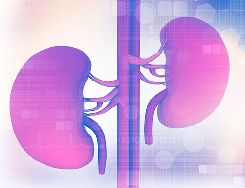
How Kidney stones are formed
Kidney stones are formed due to supersaturation of urine. This leads to crystal formation and these further aggregate to form a kidney stone.
The formation of kidney stone starts at the tip of calyces. The natural history of kidney stones is, that either they grow at the site or they pass out into the pelvi calyceal system, through the ureter into the bladder and then out through the urethra.
The kidney stone may lie silently in the kidney or it may be symptomatic in the form of a dull aching pain in the flank. When the stone passes into the ureter, the pain is usually acute, colicky, travelling from the back to the abdomen and may radiate to the genitalia (depending on the size of the stone), while the stone in the bladder normally presents as pain in the lower abdomen, as a burning sensation while passing urine or as an interrupted flow of urination. (Very often this is associated with vomiting and nausea). All above may be associated with blood in the urine.
For Consultation & Treatment
We offer affordable, highest-quality urology care with or without insurance.
Normally offered to the patients with very small kidney stones e.g. 2 to 5 mm stone lying in the peripheral (Calyceal) region which are asymptomatic and at the same time the patient has easy access to medical help. This approach still recommends life style change, dietary habits and regular checkup.
As per the natural history of the kidney stones, once they slip in the ureter, in 70% of the cases they will pass out. (depending on the size of stone, site at the time of presentation and type of stone). This expulsion of stone can be made painless and enhanced by the use of drugs that relieve the spasm of the smooth muscles of the ureter, and dilate the lower part of the ureter and urethra and thus help in painless expulsion of the stone. For this modality of treatment,the selection of cases is important by an experienced Urologist. Various co-morbidities must be looked into before the patient is prescribed this treatment (e.g. Diabetes, infection, hypertension etc.)
- One might require multiple sittings of lithotripsy for kidney stone or ureteric stone.
- One may require a stent insertion or auxiliary process, at times, to achieve stone-free status.
- The treatment may have side-effects and complications which the patient must be aware of at the time of treatment.
Retrograde Intra Renal Surgery includes the use of a Flexible Ureteroscope, that can be passed into the kidney through the urethra, bladder and then the ureter. The kidney stone is either removed intact using a grasping device or fragmented or dusted using HOL-YAG LASER. By this method we can even remove small kidney stones of 3 or 5 mm. Normally, in these cases, an indwelling stent is inserted for a period of 7 days to 1 month.
In this procedure, a semi-rigid (endoscope) ureteroscope is passed under vision upto the stone in the ureter, and the stone is removed intact or by fragmentation with an energy source like LASER or LITHOCLAST. Normally these patients are put on an indwelling stent to avoid pain and the complication of passage of a small fragment or oedema caused secondary to ureteroscopy.
Percutaneous Nephrolithotomy(PCNL), as evident from the name involves a tract formed artificially from the skin upto the kidney’s Pelvic-Calyceal system which normally contains the kidney stone. This tract is maintained by a hollow tube through which the Endoscope is passed into the kidney and the kidney stone is fragmented with an energy source and removed in chips.
These are the latest variants of PCNL, in which the size of the tract has decreased significantly and thus recovery is faster, less painful and with lesser complications. These methods have literally replaced conventional PCNL at KUC. These are the latest method of kidney.
Laparoscopic Stone Removal is used in case of a very large kidney stone in the ureter or the pelvis or in cases where reconstruction of pelvicalyceal system is required along with stone removal.
Reccurent Kidney Stone Disease and Metabolic Work Up
Recurrence of kidney stone is as high as 50% in 5 years of the first kidney stone presentation . There is an increase in prevalence of kidney stone disease globally. It may be attributed to changing affluent dietary habits, increased prevalence of diabetes and obesity, migration from cooler rural setting to urban area and global warming.
Evaluation of kidney stone formers includes: careful medical history, social and family history, dietary evaluation, occupation and laboratory evaluation.
Kidney stones are known to increase with obesity, hypertension and are a harbinger for Diabetes.
Laboratory investigation for all stone formers are:

Laboratory Evaluation Of Kidney Stones Nephrolithiasis:
Stone composition:
- By x-ray crystallography or infrared spectroscopy
Serum chemistry:
- Calcium
- Chloride
- Bicarbonate
- Sodium
- Potassium
- Phosphorous
- Blood urea nitrogen
- Intract parathyroid hormone(if high normal to high secrum calcium)
- 25-hydroxy-vitamin D(if low urine calcium or serum calcium)
- Uric acid
Urinalysis:
- Microscopic
- pH
- Specific gravity
- Protein
24-hour urine analysis:
- For patients with recurrent stones
- Some first-time stone formers
Role Of Diet in Kidney Stones
If some abnormality is picked up in the metabolic evaluation of kidney stone patient then specific diets as prescribed by the nutrinitional dietian are required.
If no abnormality is picked up in the metabolic evaluation of the kidney stone patient, the instructions are:
- To drink enough fluid in a day so as to aim for a urinary output of 2.5 to 3 liters in 24hrs.
- To eat more than five serving of fruits and vegetables daily.
- Eat Balanced meals and maintain appropriate weight.
A kidney stone patient should be aware of certain facts
- Low fluid intake causes high urinary super saturation.
- Excessive salt intake increases the calcium in urine and decreases the oxalates.
- Excessive refined, carbohydrate, caffeine or alcohol intake increases the calcium in the urine and alcoholics also have increased uric acid.
- Low intake of fruits and vegetables decreases citrate in the urine and produces acidic urine more conducive for stone formation.
